
Utilizing state-of-the-art brain imaging technology, I seek to understand how the brain encodes, consolidates, and retrieves information it encounters
Unravelling the mysteries of memory and the brain
Hippocampal-cortical interactions support memory formation
The hippocampus is thought to index experiences represented in our neocortex in service of encoding.
Recent work has shown that the hippocampus increases its activity at “event boundaries”—the end of events that are often associated with changes in predictability.
Contemporary theories have suggested that this activity may related to encoding and if that is the case, we would expect that communication between the hippocampus and neocortex would relate to subsequent event memory.
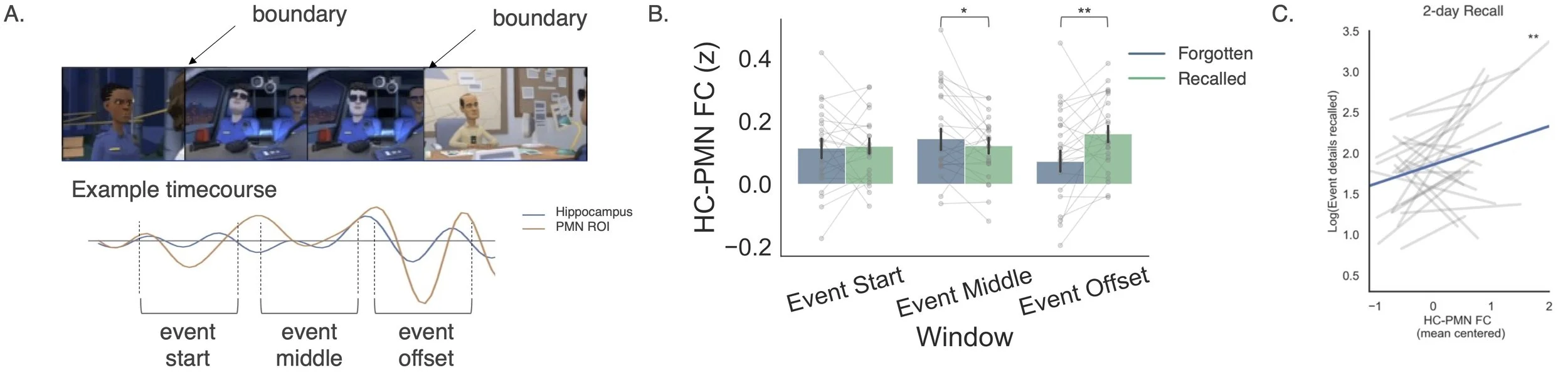
We investigated whether communication between the hippocampus and memory networks in the brain at event boundaries related to subsequent recall of the events that just finished.
In Barnett et al. (2023), we had participants watch two 15-minute cartoon movies during fMRI scanning. During encoding, calculated the functional connectivity between the hippocampus and regions of the posterior medial network (PMN) at the start, middle, and offset of each event in the movies. Participants recalled the movies in as much detail as they could into scanner-safe microphones during fMRI scanning. We then examined whether subsequent memory for the events in the movie was related to the functional connectivity between the hippocampus and PMN and discovered that events that were successfully recalled had higher connectivity between the hippocampus and PMN at event boundaries. Further, higher connectivity at boundaries during encoding was also related to the number of details participants produced when recalling an event after a 2-day delay, suggesting that offset connectivity is associated with robust encoding of rich memory indices.
Organization of cortico-hippocampal networks
The hippocampus is a critical structure in the brain that forms new memories.
It connects to a broad set of regions in the brain that are thought to contribute to this process.
I wanted to characterize exactly how these regions are functionally organized and whether that organization provides insight into how memories are formed and retrieved
In our article Barnett et al (2021), we used resting-state functional connectivity to examine network organization in the brain. We identified subgroups of regions that formed networks of communities in which regions within a network had preferential connection to other regions in the same network. A subset of these networks showed reliable connections with the hippocampus, had distinct responses during a memory-based decision making task (using a separate dataset), and were associated with partially distinct cognitive processes in an automated, data-driven meta-analysis. These findings suggest that multiple networks of distinct processing interact with the hippocampus and these interactions may subserve encoding and retrieval of events in our lives.

Hippocampal-Cortical Networks in Temporal Lobe Epilepsy
The hippocampus is often the site of seizure generation in temporal lobe epilepsy (TLE)
These seizures spread throughout the brain and may have a lasting impact on how the brain communicates
Changes in communication may contribute to common memory complaints in this population
In Barnett, Man & McAndrews (2019) we used resting functional connectivity to segment the hippocampus into its anterior and posterior components using k-means clustering in people with temporal lobe epilepsy, and also with a group of healthy control participants. While segmentation was successful in both people with epilepsy and controls, people with epilepsy had reduced connectivity to the posterior cingulate/precuneus, and medial prefrontal cortex — primary hubs of the default mode network. What’s more, those who had the weakest connections to these hubs, also had poorer memory ability.
Brain Network Properties and Cognition in Temporal Lobe Epilepsy
My PhD work focused largely on understanding how brain activity and structure relates to language and memory in people with temporal lobe epilepsy (TLE).
For some people with TLE, seizure activity can result in poorer than normal memory and naming ability.
Surgery to remove the seizure generating tissue is often an effective treatment when medication fails to control seizures, but can sometimes lead to further deficits.
Much of my work was directed at understanding the brain patterns related to these cognitive abilities and finding which patterns were related to greater risk following surgery.
Following surgery in the language dominant temporal lobe, a subset of people will experience a deficit in their naming ability. In Audrain, Barnett & McAndrews (2018), we found that the pattern of language network connectivity at rest preoperatively was related to naming ability following surgery. Specifically, people with abnormal patterns of network connectivity, compared to a healthy control template network, were at greatest risk of postoperative naming decline. By examining these networks in patients, clinicians may be able to provide more informative information and counselling for surgical candidates


Further reading on this topic:
The Hippocampus and Context
The hippocampus is a critical brain structure for forming memories.
The cells here track where an organism is in space and are also sensitive to temporal information.
These sensitivities are thought to reflect some sort of mapping that allows for memory encoding and retrieval of context and events.
In Barnett et al. (2014), we examined whether the hippocampus was sensitive not only to temporal order information, but also temporal duration information.
We showed participants a sequence of 4 scenes, which we call events, and instructed participants to remember the sequences of these events. Each of the 4 events was displayed for a different duration of time. Following a brief delay of 3.5 seconds, we showed them the same 4 events again. This time when they saw the events, we had either changed the presentation order of the events, changed the duration of time each event was presented, changed the duration of the intervals in between the events, or left the events completely the same. The participants were asked to indicate whether the sequence had changed in ANY way or whether it was the same.
The participants were very accurate at detecting changes, though they were most accurate for detecting changes in the presentation order. When a mismatch was detected (compared to match) in the sequence of the events, the hippocampus showed an increase in activity. When a mismatch was detected for the duration information, the hippocampus showed a depression of activity.
During these duration mismatches, the hippocampus was showing increased connectivity to timing regions such as the cerebellum, caudate and pre-SMA, but also to areas thought to represent context, like the posterior cingulate cortex.
Thus, the hippocampus is sensitive to both order and duration, which may help it in its role to map contexts for memory encoding and retrieval.

ESM-M: event sequence mismatch
ESM: event sequence match
IDM-M: interval duration mismatch
IDM: interval duration match
EDM-M: event duration mismatch
EDM: event duration match

Red circles indicate timing regions, yellow circle indicates context region


